Collegedunia Team Content Curator
Content Curator
Cardiac muscle (or myocardium) forms the thick middle layer of the heart. The three types of muscles found in the body are cardiac muscles, skeletal, and smooth muscles. The myocardium is surrounded by a thin outer layer called the epicardium and an inner endocardium.
Cardiac muscle contracts rhythmically and is not controlled voluntarily; thus, making it different from the functioning of skeletal muscles. One of the three main muscle types that are only present in the heart is cardiac muscle, also known as myocardium in vertebrates.
| Table of Content |
Key Terms: Cardiac Muscles, Muscle Cells, Heart, Fibrillation, Cardiac, Fibers, Muscle Organization
Cardiac Muscle Structure
[Click Here for Sample Questions]
Here, let's take a closer look at the structure and function of the cardiac muscles.
Detailed Anatomy
Human heart is majorly made of cardiac muscle, commonly known as the myocardium. The outside epicardium also called the visceral pericardium, and the inner endocardium is joined to create the heart wall by a substantial layer of the myocardium.
The endothelium that lines the blood veins that attach to the heart lines the cardiac chambers, which cover the cardiac joints and valves. While the epicardium, a component of the pericardium—the sac that envelops, protects and lubricates the heart—can be found on the myocardium's periphery.
Cardiac Muscle Cells
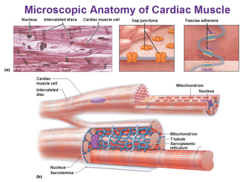
Cardiomyocytes, also known as cardiac muscle cells, are the pumping cells that enable the heart to beat. To effectively pump blood from the heart, each cardiomyocyte must contract in concert with its neighboring cells, creating what is known as a functional syncytium. The heart may not pump at all if this coordination fails, even when individual cells are contracting, as can happen with irregular heart rhythms like ventricular fibrillation.

Cardiac Muscle Anatomy
T-Tubules
[Click Here for Sample Questions]
The tiny tubes known as T-tubules extend from the cell's surface to its inside. These share a phospholipid bilayer structure and are one continuous membrane with the cell membrane. They are open at the cell's exterior, or extracellular fluid surface.
T-tubules in cardiac muscle are fewer in number but wider and larger than those in skeletal muscle. They form a transverse-axial network in the center of the cell by running into and beside it. They are located close to the sarcoplasmic reticulum, the cell's internal calcium storage. In a dyad combination, a solitary tubule is paired with a terminal cisterna from the sarcoplasmic reticulum.
Discs with IntercalationThe cardiac syncytium is a network of cardiomyocytes connected by intercalated discs that enable the syncytium to participate in the synchronized contraction of the heart, allowing for the rapid transmission of electrical impulses across a network. There are cardiac connecting fibers that join the ventricular and atrial syncytia. |
What Distinguishes Skeletal Muscle from Cardiac Muscle?
[Click Here for Sample Questions]
The body is moved by the contraction of skeletal muscles, which are affixed to bones. Heart muscles called cardiac muscles contract to pump blood.
Between 10 and 100 micrometers broad, skeletal muscle fibers are long and thin. With a width of 20 to 200 micrometers, cardiac muscle fibers are thicker. Unlike the heart muscle, skeletal muscle is also structured in bundles called fascicles.
When the heart needs to fill with blood, the branching architecture of the cardiac muscles permits them to do so (unlike skeletal muscles).
Also check:
Cardiac Muscle Organization
[Click Here for Sample Questions]
Because it has a considerably more intricate structure than skeletal muscle, cardiac muscle is significantly distinct from the latter. In contrast to skeletal muscles, which can only contract, cardiac muscle may both contract and relax.
As a result, heart tissue will be far more atypically shaped than skeletal muscles. The sarcoplasmic reticulum, which stores the calcium ions required for cell contraction, is connected to T tubules found in cardiac muscle cells.
The heart and the tissues connected to it are surrounded and safeguarded by the myocardium, a thin yet sturdy membrane. The myocardium, endocardium, and epicardium make up the heart wall.
All four of the heart's chambers are lined on the inside by the endocardium, which also covers the heart valves. The outside is covered by a thin coating of epicardium.
Cardiac Muscle Disease Clinical Implications
[Click Here for Sample Questions]
A collection of conditions known as cardiac myopathic illnesses have an impact on how well the heart can function. There are some of these illnesses that, if caught early enough, can be cured, but there are also some fatal diagnoses.
Cardiovascular disease refers to any chronic condition affecting the heart or blood vessels, such as those affecting the heart muscle, valves, or arteries. Congestive heart failure, hypertension, and other conditions are included in this phrase.
Diseases related to defects in the structure of the cardiopulmonary system or congenital abnormalities are often chronic. They range from common conditions like coronary artery disease to rare genetic diseases that affect many organs. Some examples include atrial septal defect (ASD) and patent ductus arteriosus (PDA).

Cardiac Muscle Disease Clinical Implications
Things to Remember
- Cardiac muscle regulates the contractility of the heart and the pumping action. The cardiac muscle must contract with enough force and enough blood to supply the metabolic demands of the entire body.
- Cardiac muscle tissue cells characteristics: involuntary and intrinsically regulated, branched, striated, and single nucleated.
- Cardiac muscle cells form a branched cellular network in the heart.
- Cardiac muscles are connected by intercalated disks and are organized into layers of myocardial tissue wrapped around the chambers of the heart.
- Two types of cardiac muscle fibers: myocardial contractile cells and myocardial conducting cells (pacemaker cells).
Also check:
Sample Questions
Ques 1. What are cardiac muscles? What are its functions? (2 Marks)
Ans. Cardiac muscles are a type of specialized, striated muscle found only in the heart. They are under the control of the autonomic nervous system, which means they are involuntary and work autonomously.
Cardiac muscles consist mainly of cells called cardiomyocytes which are responsible for the generation of contractile force as well as provide structural and functional support for the cardiac muscle tissue. They also contain blood vessels that supply nutrients to cardiac muscle tissue and remove waste products. Cardiac muscles also contain nerve cells or neurons that initiate impulses.
Ques 2. Why is Cardiac Muscle Function Important? (1 Mark)
Ans. Cardiac muscle function is important to the body because it pumps blood through the body. Cardiac muscles are made up of special cells called myocytes that can contract and relax in alternate phases.
Ques 3. What is the difference between Skeletal and Cardiac Muscle? (1 Mark)
Ans. Skeletal muscles are attached to bones and they contract to move the body. Cardiac muscles are found in the heart and they contract to pump blood. Skeletal muscle fibers are long and thin. Cardiac muscle fibers are thicker.
Ques 4. How do fibroblasts function? (2 Marks)
Ans. The crucial supporting cells within the heart muscle are described as cardiac fibroblasts. Contrary to cardiomyocytes, which are able to contract the heart vigorously, they are primarily in charge of producing and maintaining the extracellular matrix, which serves as the mortar in which cardiomyocyte bricks are embedded. When responding to injuries like myocardial infarction, fibroblasts are crucial.
Ques 5. What is the Extracellular Matrix? (3 Marks)
Ans. Continuing the wall analogy, the extracellular matrix surrounds the bricks of fibroblasts and cardiomyocytes like mortars on a wall made of the heart muscle. Proteins like elastin and collagen as well as polysaccharides (sugar chains) referred to as glycosaminoglycans make up the matrix.
Together, these compounds provide the muscle cells with strength and support, maintain the hydration of the muscle cells, and increase the flexibility of the heart muscle by tying together water molecules.
Ques 6. What is the cardiac muscle's clinical importance? (2 Marks)
Ans. In many industrialized countries, diseases of the heart muscle are the main cause of death, and they have significant clinical implications. The most frequent ailment that affects the cardiac muscle and results in decreased blood flow to the heart is ischemic heart disease. In ischemic heart disease, atherosclerosis causes the coronary arteries to narrow.
Ques 7. Distinguish between the atria and the ventricles. (2 Marks)
Ans. The heart's atria and ventricles are created by cardiac muscle. There are a few variances even if the muscular tissue in the heart chambers is the same. The myocardium in the ventricles is thick, whereas the myocardium in the atria is thinner, allowing the heart to contract forcefully. The myocardium, which is made up of individual myocytes, differs between the cardiac chambers as well. The T-tubule network is denser and larger and longer in ventricular cardiomyocytes.
Check-Out:


Comments