


Content Writer-SME
The human ear is a complex and remarkable organ responsible for the sense of hearing and, to some extent, the sense of balance. It is an intricate system that allows us to perceive a wide range of sounds and helps us navigate the world around us. The process of hearing involves the transformation of mechanical vibrations (sound waves) into electrical signals that can be interpreted by the human brain.
- This intricate mechanism allows humans to perceive a vast spectrum of sounds, from the gentle rustling of leaves to the complex melodies of music.
- The hearing range of the human ear is approximately 20 Hz to 20,000 Hz. Sounds beyond this range are not detected by our ear.
- Apart from helping us hear, the human ear also helps us maintain body balance or equilibrium.
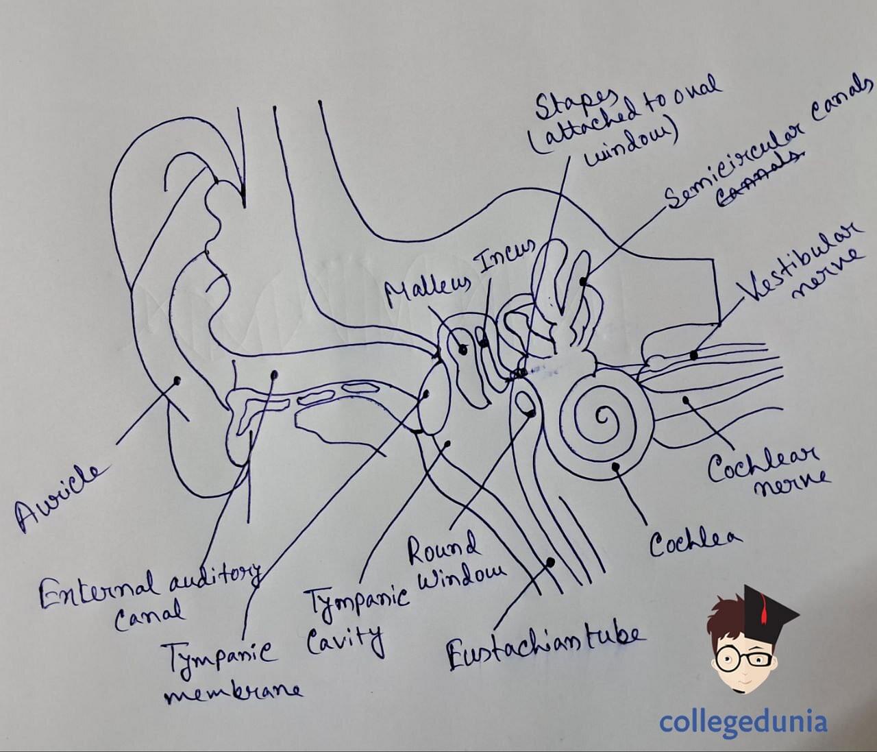
Structure of Human Ear
[Click Here for Sample Questions]
The human ear is composed of three primary divisions which are known as the outer, middle, and inner ear. The structure of the human ear is explained as follows:
Outer Ear
Sound waves are collected and directed into the ear canal via the outer ear, an external part of the human ear that is essential to hearing. The outer ear is composed of the pinna, ear canal and tympanic membrane.
- Pinna assists in capturing and channelling sound waves to the ear canal, which is a tube that leads internally.
- The tube that joins the middle ear to the outer ear is called the ear canal, sometimes referred to as the auditory canal or meatus.
- Sound waves cause the tympanic membrane to vibrate, converting it into mechanical energy that is sent to the middle ear.
Middle Ear
Sound waves cross the external part of the ear and arrive at the middle ear where the eardrum is located.
- The eardrum vibrates upon impinging sound waves.
- This vibration is conducted to the small bones called ossicles.
- These chains are made of fine bones which increase the amplitude to make it possible to transmit forwards to the inner ear.
Inner Ear
Located inside the skull is an intricate structure known as a bony labyrinth, which contains fluid in the inner ear. The labyrinth consists of the cochlea used in hearing and the vestibular system critical for balance.
Cochlea
It is also important to note that the organ of hearing is known as the cochlea and is somewhat similar to a snail shell.
- It is full of about three thousand hair cells involved in transforming mechanical vibration into an electric stimulation.
- The brain interprets these signals as sound by using the auditory nerve.
Vestibular System
Balance and spatial orientation are also maintained by the vestibule which lies beside the cochlea. It is composed of the three semicircular canals (which are attached to the otolitic organs) that sense the tilt or rotation movements as well as the linear acceleration, sending the signals to our brain for balancing purposes.
Structure of the Human Ear
Hearing Process
[Click Here for Previous Year Questions]
The mechanism by which the human ear translates sound waves into sensory information to be perceived in the cerebral cortex is indeed amazing!
- Sound Wave Reception in the Outer Ear:
It all starts with the external ear which includes both the pinna and the ear canal. The pinna is like a funnel that traps the sound waves in the surroundings and channels them through the ear canal. As these waves move in the canal they reach the eardrum which is an ultrathin dividing line between the outer region and inner zone (middle ear).
- Amplification in the Middle Ear:
The eardrum vibrates when sound waves contact it. These vibrations are then transferred onto the three tiny bones of the inner ear; the malleus, the incus, and the stapes. These chains of bones work as a lever system that augments the vibrations and sends them to the oval window, which is a membrane-coated opening into the inner ear.
- Transformation in the Inner Ear (Cochlea):
Coiled in the inner ear is the cochlea—a spiralled tube filled with fluid that looks like a snail shell. The fluid in the cochlea ripples as the enhanced vibrations get into the cochlea via the oval window. Thousands of microscopic hair cells lining the cochleas are stimulated by this movement.
- Conversion into Electrical Signals:
These cells are important in transforming vibrations into electric pulses. These hair cells produce electrical impulses when stimulated with fluid motion. The precise spot where a single hair cell is stimulated represents the frequency of the sound wave and allows the brain to distinguish between various pitches.
- Transmission to the Brain:
Once generated, the hair cell’s electrical signals traverse nerves in the form of a fibre bundle which is known as the auditory nerve to reach the brain. The fastest route of this nerve path ensures the speedy information delivery to the auditory cortex which is the part of the brain concerned with sound.
- Auditory Cortex Interpretation:
Electrical signals sent to the auditory cortex are highly processed in a complicated manner. The brain determines the frequency, the loudness, and how long each signal lasts. It allows us to understand speech, listen to music, and perceive important information in our environment.
Functions of Human Ear
[Click Here for Sample Questions]
An amazing organ consisting of the outer ear (pinna), the middle ear (including the tympanic cavity), and the cochlea of the inner ear enables us to hear and balance.
Outer Ear
The major area during the auditory experience is the external section which comprises the pinna and the ear canal. Sound waves from the surroundings are captured by it and directed toward the internal components of the human ear.
- The outer ear protects the middle and inner ear's fragile structures from injury and contaminants.
- The famous ear pinna which is sometimes overlooked, functions as a kind of funnel directing soundwaves into the canal.
- Sound waves travel through this curved path which ends with an eardrum of soft membrane, distinguishing the external and internal parts of the ear.
Middle Ear
The middle ear, which lies between the outer and inner ears, is an important component of the auditory system. This part of the human ear is an air-filled chamber that contains the hammer, anvil, and small bones of the stirrup.
- It is essential for hearing since it transmits and amplifies sound waves.
- These, in turn, vibrate and act as an amplifier for the sound waves that reach the eardrum.
- This is a ballet of sorts that converts external signals into a language that the internal ear will understand.
Inner Ear
An essential component of the human auditory and balancing system is the inner ear. It is situated well inside the skull's temporal bone. Deep inside the inner ear is the cochlea, a coiled and fluid-filled organ that looks like a spiral snail shell. It is here when auditory transduction happens.
- These vibrations are then amplified by hair cells which mimic tiny receptors.
- They convert these vibrations into electrical signals that the auditory nerve then carries to the brain.
- The detailed process that underpins our perception of sound is very complicated.
Things to Remember
- The pinna is like a funnel that traps the sound waves in the surroundings and channels them through the ear canal.
- Sound waves cross the external part of the ear and arrive at the middle ear where the eardrum is located.
- The labyrinth consists of the cochlea used in hearing and the vestibular system critical for balance.
- The inner ear comprises of two parts: A bony labyrinth and a Membranous labyrinth
- The fluid in the cochlea ripples as the enhanced vibrations get into the cochlea via the oval window.
- The cochlea is the main organ in the inner ear.
Read More:
| Related Topics | ||
|---|---|---|
| Central Nervous System | Living and Non-Living Thing | Nutrition In Human Beings |
| Circulatory System | Skeletal System | Nervous System |
Previous Year Questions
- Extraction of metal from the ore cassiterite involves...[JEE Advanced 2011]
- Commonly used vectors for human genome sequencing are...[NEET UG 2014]
- Interfascicular cambium and cork cambium are formed due to..
- Pneumotaxic centre is present in...[UP CPMT 2007]
- Reaction of HBr with propene in the presence of peroxide gives….[NEET UG 2004]
- Assuming the expression for the pressure exerted by the gas on the walls of the container, it can be shown that pressure is...[MHT CET 2016]
- Which among the following is the strongest acid?...[TS EAMCET 2017]
- Isopropyl alcohol on oxidation forms..
- A vector is not changed if..
- Which of the following arrangements does not represent the correct order of the property stated against it?...[JEE Main 2013]
Sample Questions
Ques. What does the external ear do? (2 marks)
Ans: The main function of the external ear is to focus sounds on the narrow opening to the ear. The external ear consists of the pinna and the ear canal. It is responsible for major auditory experiences for humans. The external ear protects the organs in the middle and inner ear from any damage.
Ques. What is that portion between the outer and middle ear? (2 marks)
Ans: The portion between the outer and the middle ear is called the Eardrum (Tympanic Membrane). It's a fragile, but tight construction similar to an infant trampoline. The invisible messengers of sound travel in the form of sound waves and when they reach the eardrum it gets moved. As the ripples in a calm pool, this motion initiates a spectacular cause-and-effect phenomenon.
Ques. Name the three bones in the middle ear. (2 marks)
Ans: There are three main bones in the middle ear – entering the ossicles. The malleus, incus, and stapes are the other three bones that form the ossicles. The ossicles take up the beat when the eardrum dances to the incoming sound waves. Click clack, a dance orchestrated by vibrations; hammer on the anvil, through the stirrup and a symphony starts.
Ques. What part of the body does the cochlea form? (2 marks)
Ans: The inner Ear is the mechanical version of a sound engineer transforming the amplified vibrations passing through the middle ear into electric signals for transmission by the auditory nerve to the brain. Similarly, the cochlea can be compared to a highly refined musical instrument whose hair fibres are poised to perceive even the smallest details of the sound.
Ques. What do the hair cells of the cochlea do? (2 marks)
Ans: The minute architects of the cochlea and hair cells convert mechanical vibration into electrical signals for the brain to finally perceive sound. These cells are specially adapted and look like receptors that live inside the winding spirals in the cochlea. The hair cells are very precise when passing via the external and interior ear through the cochlea in this case
Ques. Which nerve sends electrical signals from the cochlea to the brain? (2 marks)
Ans: The auditory nerve functions to relay the auditory information from the cochlea to the brain. It is the sole messenger that transmits the electrical impulses produced in the cochlea to the brain for decoding. The complex hair cells of the cochlea convert vibration-based sound into electric pulses which are sent via the auditory nerve towards the brain’s auditory processing centers.
Ques. Where in the brain does one process sound? (2 marks)
Ans: The processing of sound takes place mainly in the auditory cortex. This section lies in the temporal lobe and is capable of translating the impulses received from the auditory nerve. The signals go through the thalamus before reaching the auditory cortex which decodes the pitches, volume, and other auditory features.
Ques. What is the major function of the vestibular system in the internal ear? (2 marks)
Ans: The main function of the vestibular system in the internal ear is to maintain balance and spatial orientation. This delicate system identifies the change in head movement, thus passing all the necessary information to the brain in the form of signals and the brain will act accordingly.
Ques. Which part of the ear is reflexively contracted due to a high-volume sound to dampen vibration? (2 marks)
Ans: Middle Ear Muscles contract due to a high-volume sound to dampen the vibration perceived by the ear. The reason behind this contraction is to prevent damage to the inner ear membranes and the organs. This contraction allows the high-volume sound to slow down and then reach the inner ear.
Ques. What is the coiled tube filled with fluid that lies inside the ears used in hearing? (2 marks)
Ans: Cochlea is the tube filled with the fluid that lies inside the ears. Mechanical vibrations are converted into electrical signals within the cochlea. Following this, the auditory nerve is responsible for transferring the signals further to the brain. And, then the brain will send messages through neurons. The cochlea is an essential part of the ear used for hearing.
For Latest Updates on Upcoming Board Exams, Click Here: https://t.me/class_10_12_board_updates
Check-Out:


Comments